Unlike mammals, some vertebrates like salamanders and zebrafish are known for their remarkable capacity to regenerate complex tissues, such as limbs, tails, and tailfins. Dissecting the fundamental mechanisms that enable these animals to undergo robust regeneration may be instructive for applications in regenerative medicine.
One of the most puzzling features of tissue regeneration in these animal models is the exact reproduction of missing parts upon completion of the process. Many scientists have attempted to examine the limits of this fidelity. For instance, a study has reported that 27 consecutive amputations of zebrafish tailfin over an 11-month period cannot affect the size of the regenerated tailfin. Similarly, the extent to which adult salamanders are able to regenerate eye structures was put to an extreme test; after 18 consecutive lens-removal surgeries over 16 years, scientists have reported that no structural or molecular abnormalities could be observed in regenerated lens tissues. Since Spallanzani conducted the first repetitive amputation experiments on salamander limbs and tails over 250 years, little has been learned about how anatomical details, the so-called positional information, are ‘memorized’ to instruct the growth of a perfect replacement. Scientists do not know where do these animals store ‘positional memory’? How can the memory be effectively decoded and retrieved? Is it possible to rewrite the memory?
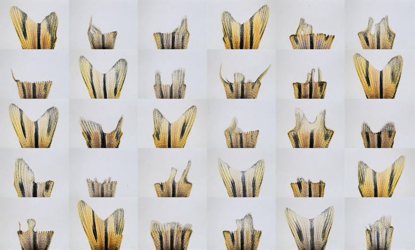
From a chemical mutagenesis screen, a research team led by Dr. Chen-Hui Chen at the Institute of Cellular and Organismic Biology, Academia Sinica (Taiwan) accidentally identified a novel zebrafish mutant with defects in positional memory. They found the mutation can cause tailfins to regenerate with high variability in sizes and shapes. Based on high-resolution genetic mapping and complementation assays, they determined DNA polymerase alpha subunit 2 (pola2) is the mutated gene. The gene activity has a direct impact on blastemal proliferation and size. Through manipulating pola2 activity, they developed an approach to effectively rewrite positional memory. The new memory is able to direct growth of tailfin and scales, even after repetitive injuries. Through collaboration with Drs. Jr-Kai Yu and Yi-Hsien Su at the Institute of Cellular and Organismic Biology, they found similar effects upon transient pharmacological disruption of progenitor cell proliferation after head or tail amputation in amphioxus and annelids. There findings suggest that the regulatory mechanisms identified in this study may be evolutionarily conserved across vertebrate and invertebrate species.
Taken together, this study discovered a key genetic factor for altering positional memory in zebrafish tissues and established a working model in evolutionarily distant animals. The importance of these findings is that they provide the first means to alter the fidelity of positional memory. Thus, classic regeneration models that assume the memory is unmalleable may need revisiting and refinement. This study entitled “Genetic reprogramming of positional memory in a regenerating appendage” will be published in the incoming issue of Current Biology (2019).