The 2014 Nobel Prize in Chemistry was awarded to three scientists who have advanced the optical microscopy system beyond the limits of optical resolution. Assistant Research Fellow Bi-Chang Chen, from the Academia Sinica Applied Science Research Center, and Academician Ann-Shyn Chiang, Dean of the Department of Life Sciences and Brain Research Center of Tsing Hua University, jointly developed a new generation of three-dimensional optical super-resolution microscopy – Microscopes allow super resolution imaging from the cell level to the tissue level, which deconstructing the dopaminergic neural network of the whole brain of Drosophila, and seeing the regeneration of memory proteins at specific neuronal synapses. The results of this research have been published in Nature Communications on October 18, 2019.
Before coming back to the Academia Sinica, Bi-Chang Chen worked in Dr. Eric Betzig’s lab, one of the winners of the 2014 Nobel Prize in Chemistry. Recently, he cooperates with Academician Ann-Shyn Chiang to make biological tissues transparent under the microscope, which enables locating and quantifying the spatial distribution of each single protein. This technology is expected to bring breakthroughs in tissue physiology and pathology research and has the potential to uncover the mystery of the brain’s memory mechanism.
Bi-Chang Chen said that this research results can be regarded as an advanced version of his research in Communications Biology earlier this year. The study used a lightsheet localization microscope to achieve a three-dimensional spatial resolution of less than 100 nm, and the size of the nuclear pores was visible.

Fig. 1: Left: Super-resolved whole brain dopaminergic neurons. Middle: Single dopaminergic neuron located in fly eye can be manually segmented. Right: VMAT distribution in whole DPM neuron can be located.
With the foundation of Bi-Chang Chen’s technology, Academician Ann-Shyn Chiang hopes to use it to super-resolve the complete Drosophila brain, that is, to locate all protein molecules in a tissue nearly 10,000 times larger than a single cell. Chiang’s vision is challenging for Bi-Chang Chen, who has a background in optical technology because if he wants to get Dr. Eric Betzig’s work for the Nobel Prize in Chemistry, he will face various difficulties to apply two-dimensional cell super-resolution imaging to three-dimensional tissue research. For example, the opaque Drosophila brain has low visible light transmittance, is difficult to evenly distribute fluorescent dyes, and how to use a lightsheet microscope for observing very thin monolayer cells to three-dimensional Drosophila brain.
To solve these problems, laboratories in various fields must cooperate. First of all, the whole brain tissue clearing method invented by Academician Ann-Shyn Chiang can make the Drosophila brain transparent within minutes and allow visible light to penetrate the whole brain. Prof. Yeu-Kuang Hwu from the Institute of Physics of Academia Sinica has modified the grafting of fluorescent molecules; Academician Ting-Kuo Lee also gave guidance on image processing; Prof. Peiling Chen from Appreciation Center had experience in processing images of super-resolution images; Associate Researcher Shuwei Chang participated in the simulation of optical systems. Finally, Dr. Li-An Chu and Dr. Chien-Han Lu, carried out all the experiments and analysis and finally let the lightsheet localization microscope super-resolve the distribution of protein molecules in the whole brain.
To resolve super-resolution imaging of two-dimensional cell images usually took one night in the past. With the new microscopy and image analysis system developed by Yen-Ting Liu, a talent undergraduate student from National Taiwan University, can analyze the molecular image of a whole fruit fly in one day, therefore this tool can be used to Statistical analysis of the number of proteins in any neuron in any part of Drosophila brain.
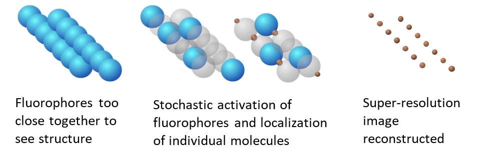
Fig. 2: Principle of localization microscopy
Also, the paper discusses the distribution of vesicular monoamine transporter in the dorsal paired medial neurons in the fruit fly. By comparing the brains of Drosophila with memory training and no memory training, the team found that only some of the synapses had protein increases, which means that in a single nerve cell, memory not only exists in the cell body but also stores at the bridges where nerve cells communicate.
This project can be realized via several funding agency. In addition to the internal funding support of the Applied Science Research Center, also including a number of grants: the Academia Sinica’s grant for forward-looking infrastructure development, the Higher Education Sprout Project funded by the Ministry of Science and Technology and Ministry of Education in Taiwan. The authors of this research project including Li-An Chu, Chieh-Han Lu, Shun-Min Yang, Yen-Ting Liu, Kuan-Lin Feng, Yun-Chi Tsai, Wei-Kun Chang, Wen-Cheng Wang, Shu-Wei Chang, Peilin Chen, Ting-Kuo Lee, Yeu-Kuang Hwu, Ann-Shyn Chiang & Bi-Chang Chen. Publication link: https://www.nature.com/articles/s41467-019-12715-3
In addition, in terms of talent cultivation, Shangjr Guo, director of the Center for Applied Science in Academia Sinica, said that the center and Department of Life Science and Brain Research Center in Tsing Hua University are about to sign a memorandum of cooperation on the doctoral class of Elite, hoping to expand cooperation on the basis of the existing units. The program will provide monthly scholarships for doctoral students with excellent academic performance and research potential to jointly cultivate more basic scientific research talents.